Compact Bone Diagram Endosteum - Bone Tissue Amboss : Flat bones, like those of the cranium, consist.
byTed Mcknight-
0
Compact Bone Diagram Endosteum - Bone Tissue Amboss : Flat bones, like those of the cranium, consist.. These cells resemble bone lining cells in both the endosteum and periosteum. In this type of bone, the lamellae are organised into concentric circles, which surround a vertical haversian in both types of bone, the external surface is covered by a layer of connective tissue, known as the periosteum. The _____ covers all bones except parts of joints enclosed with a joint capsule. Spongy osseous tissue has similar structures. It is a thin covering that surrounds the.
Usually bones that are thin and curved. It acts as a coating for the inner compact bone and the trabeculae of the spongy tissue. Definition and functions the endosteum is a structure in the middle of bone tissue and bone marrow. Moreover, periosteum and endosteum cover the compact bone from outside and inner surface respectively. The endosteum is thin connective.
Anatomy Of A Bone Coloring from www.biologycorner.com These blood vessels transport nutrients to nourish the endosteum. Sclerostin inhibits bone formation mostly by antagonizing lrp5/6, thus inhibiting wnt signaling. Endosteum also has few connective tissues fibers and blood vessels. The osteoprogenitor cells of the preosteoblasts present in this connective tissue lining, differentiate into. It covers the loose structures found inside the bone. In this type of bone, the lamellae are organised into concentric circles, which surround a vertical haversian in both types of bone, the external surface is covered by a layer of connective tissue, known as the periosteum. Structure of compact bone, spongy bone, periosteum, and endosteum. These cells add the compact bone to the bony callus to form a bone tissue that is similar to the original, normal bone.
A central tube called a periosteum, the equivalent to endosteum on the outside of the bone, plays a vital role in the healing of fractures.
These cells add the compact bone to the bony callus to form a bone tissue that is similar to the original, normal bone. Endosteum is a thin, soft, connective tissue, lining the cavity of long bones like humerus and femur. Structure of compact bone, spongy bone, periosteum, and endosteum. A central tube called a periosteum, the equivalent to endosteum on the outside of the bone, plays a vital role in the healing of fractures. What is the difference between compact bone and cancellous bone?
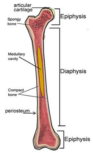
Notes Ch 7 Skeleton from www.biologycorner.com Also called cortical bone, the compact variety usually features a haversian system, or cylindrical unit within the structure. Osteocytes synthesize bone and reside on the surfaces of bone: Compact bone, also called cortical bone, is the hard, stiff, smooth, thin, white bone tissue that surrounds all bones in the human body. Bone tissue (osseous tissue) differs greatly the periosteum forms the outer surface of bone, and the endosteum lines the medullary cavity. A central tube called a periosteum, the equivalent to endosteum on the outside of the bone, plays a vital role in the healing of fractures. Sclerostin inhibits bone formation mostly by antagonizing lrp5/6, thus inhibiting wnt signaling. ¨ an osteon (or haversian system): To know the architecture of compact and spongy (cancellous) bone.
¨ an osteon (or haversian system):
These are mostly compacted bone with little marrow and include most of the bones in the limbs. It is a thin covering that surrounds the. These cells resemble bone lining cells in both the endosteum and periosteum. The inset shows the lamellae of the compacta. This endosteal surface is usually resorbed during long periods of malnutrition, resulting in less cortical thickness. It is a thin covering that surrounds the. A similar layer, the endosteum. The densest and strongest bones in the body. To know the architecture of compact and spongy (cancellous) bone. To know the structures of a synovial joint and a symphysis joint (intervertebral disc). Flat bones, like those of the cranium, consist. It is made up of. Sclerostin inhibits bone formation mostly by antagonizing lrp5/6, thus inhibiting wnt signaling.
The basic units of compact bone are called osteons or haversian systems. Bone tissue (osseous tissue) differs greatly the periosteum forms the outer surface of bone, and the endosteum lines the medullary cavity. This endosteal surface is usually resorbed during long periods of malnutrition, resulting in less cortical thickness. It is a thin covering that surrounds the. Are located in the periosteum and endosteum.
Sclerostin inhibits bone formation mostly by antagonizing lrp5/6, thus inhibiting wnt signaling. In this type of bone, the lamellae are organised into concentric circles, which surround a vertical haversian in both types of bone, the external surface is covered by a layer of connective tissue, known as the periosteum. The endosteum is located on the internal surface of the bone, being the membranous layer that covers the medullary cavity, the bony trabeculae (spongy part of the bone), the haversian canals and internal walls of the compact long bones. This process can take several months. What is the difference between compact bone and cancellous bone? Are located in the periosteum and endosteum. They are very difficult to distinguish from the surrounding connective tissue cells. The basic units of compact bone are called osteons or haversian systems. These are mostly compacted bone with little marrow and include most of the bones in the limbs. Flat bones, like those of the cranium, consist. It is a thin covering that surrounds the. Describe how bones are nourished and innervated. The endosteum is thin connective.
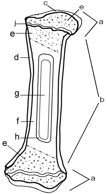
The endosteum is located on the internal surface of the bone, being the membranous layer that covers the medullary cavity, the bony trabeculae (spongy part of the bone), the haversian canals and internal walls of the compact long bones compact bone diagram. The densest and strongest bones in the body.